Why Choose an Optical Laryngoscope for Better Intubation?
Choosing the right laryngoscope can influence the clarity, speed, and safety of airway management. An Optical Laryngoscope provides an illuminated view through an integrated camera or optical pathway. This design may help clinicians visualize the glottis when direct line-of-sight techniques prove difficult.
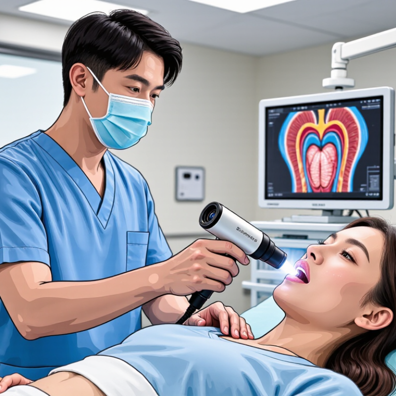
In practice, the difference can be visible immediately. A monitor may show the epiglottis, vocal cords, and surrounding anatomy in greater detail. This shared view can support team communication during intubation. It may also assist teaching, documentation, and careful review after the procedure. However, better visualization does not guarantee successful intubation. Patient anatomy, secretions, blood, fogging, positioning, and operator experience still matter.
Experienced airway professionals use optical devices within a structured assessment, not as a universal solution. Training should include familiarization with the handle, blade shape, screen position, and cleaning process. Small delays can occur while adjusting the camera or clearing the lens. That detail deserves attention.
Clinical guidance and device instructions should inform equipment selection. Evidence can support confidence, but local protocols and patient-specific risks remain essential. An Optical Laryngoscope may offer meaningful advantages in selected cases, especially when traditional views are limited. Its value depends on preparation, technical skill, reliable equipment, and sound clinical judgment. Even advanced technology has boundaries. Recognizing those boundaries is part of safer airway practice.
Optical Laryngoscopes: Imaging Principles and the Cormack–Lehane Scale
Why Choose an Optical Laryngoscope for Better Intubation?
An optical laryngoscope uses a distal lens, prism, or image pathway to transmit the glottic view. It reduces the need to align the oral, pharyngeal, and laryngeal axes. The operator watches the epiglottis and vocal cords through an eyepiece or screen. Small movements matter. A slight blade adjustment can change the view dramatically.
The Cormack–Lehane scale supports structured assessment during intubation. Grade I shows the full glottis, while Grade II shows only part of it. Grade III reveals the epiglottis without visible vocal cords. Grade IV shows neither the glottis nor the epiglottis. Modified grading can separate IIa from IIb, although interobserver agreement is not perfect. That limitation deserves attention.
The UK Fourth National Audit Project reported major airway complications at approximately 1 in 22,000 anesthetics. Its findings underline the need for reliable visualization and early rescue planning. A Cochrane review found that videolaryngoscopy generally reduces failed intubation compared with direct laryngoscopy. However, those findings should not be transferred automatically to every optical device. Blade geometry, lens contamination, secretions, patient anatomy, and operator training still influence success. A clear image is not the same as a successful tube passage. Practice should include identifying the Cormack–Lehane grade, limiting repeated attempts, and confirming tracheal placement with capnography. Errors remain possible. That is the uncomfortable part.
First-Pass Success: Cochrane Evidence for Video Laryngoscopy
Why Choose an Optical Laryngoscope for Better Intubation?
First-pass success matters when oxygen reserves are limited and every attempt increases airway trauma. A 2022 Cochrane Review assessed 222 studies involving 26,149 adults. Compared with direct laryngoscopy, video laryngoscopy reduced failed intubation risk, with a reported risk ratio of 0.41 (95% confidence interval, 0.30–0.55). The review also found slightly higher first-attempt success, with a risk ratio of 1.05 (95% confidence interval, 1.01–1.10).
The screen can show the glottic opening while the operator controls the tube near the patient’s mouth. That shared view helps assistants anticipate tube direction and respond earlier to a poor angle. In a crowded operating room, this visibility is practical. It is not magic. Secretions, fogging, blood, or limited mouth opening can still obscure the image. The 2022 Cochrane evidence also included varied devices, clinicians, and clinical settings, so results may not transfer perfectly to every airway.
Training remains decisive. A 2022 Difficult Airway Society guideline update supports video laryngoscopy as an important option when appropriate expertise and equipment are available. Operators should check the blade, screen, suction, and backup plan before induction. One missed detail can erase the advantage. Real-world audits should track first-pass success, oxygen desaturation, dental injury, and time to intubation, rather than relying on a clear view alone.
Why Choose an Optical Laryngoscope for Better Intubation?
First-Pass Success: Cochrane Evidence for Video Laryngoscopy
Compared with direct laryngoscopy, video laryngoscopy was associated with a modest increase in successful first-attempt intubation and a lower risk of failed intubation.
Relative risks are from a Cochrane systematic review of 64 studies involving 7,044 adult participants. A relative risk above 1 favors video laryngoscopy for first-pass success; below 1 favors video laryngoscopy for failed intubation.
Glottic Exposure: POGO Scores and Grade I–II View Rates
An optical laryngoscope can improve intubation by showing airway structures through an integrated visual pathway. Its value is clearest when glottic exposure is measured, not simply assumed. The POGO score describes how much of the vocal opening is visible. A higher percentage usually means more anatomical detail for guiding the tube. Grade I–II views also indicate useful exposure, although grading can vary between clinicians.
In clinical training, a broader POGO view may reduce repeated blade adjustments and unnecessary force. The operator can watch the tube pass toward the glottis while maintaining attention on the screen. Small movements matter. A few millimeters can change the view. However, an optical system does not guarantee a perfect laryngoscopy. Blood, secretions, fogging, poor lighting, or an unfamiliar screen angle can reduce visibility.
Reliable assessment needs more than one successful attempt. Record the POGO percentage, Cormack–Lehane grade, number of attempts, and time to view. These details reveal whether improved exposure is consistent. I would also question a high score when tube delivery remains difficult. A wide glottic image can still hide problems with tube angle, patient positioning, or operator coordination. That limitation deserves honest review.
Safety Outcomes: ASA Guidance on Attempts, Trauma, and Hypoxemia
An optical laryngoscope can provide a clearer view of the glottis during difficult intubation. Its camera may help clinicians work around limited alignment, restricted neck movement, or a crowded oral cavity. However, better visualization does not guarantee safe placement. Preparation still matters.
ASA guidance emphasizes limiting intubation attempts. Each attempt can cause bleeding, swelling, dental injury, or worsening visualization. Repeated forceful attempts may also convert a manageable airway into a critical one. Set an attempt limit before starting. The exact limit should follow clinical judgment, team experience, and local policy. Change the approach early when progress stops.
Oxygenation must remain central. Preoxygenation should be checked, not assumed. In selected patients, nasal oxygen or noninvasive ventilation may help maintain oxygen levels during airway management. An optical view can reduce unnecessary manipulation, but it cannot prevent hypoxemia caused by delay, poor mask seal, or exhausted oxygen reserves. Watch the monitor continuously. Pause when saturation falls.
Small details matter. Position the patient carefully. Suction secretions early. Confirm tube placement with waveform capnography. A useful reflection is uncomfortable: a familiar technique may feel safer than it actually is. Optical equipment can improve decision-making, yet overconfidence may create new delays. The safest team recognizes when to stop, oxygenate, call for help, and use an alternative airway plan.
Device Selection: Blade Design, Training, and Evidence-Based Metrics
Why Choose an Optical Laryngoscope for Better Intubation?
Device selection should begin with blade design, not screen size. A Macintosh-style blade supports familiar lifting mechanics and may suit routine airways. A hyperangulated blade can improve the view when alignment is difficult, but it often requires a shaped stylet. Tube delivery may become harder. Small changes matter: blade width, tip curvature, camera position, and anti-fog performance can affect control in a wet airway.
Training must match the chosen design. The Difficult Airway Society recommends structured preparation, oxygenation, limited attempts, and early escalation. The 2022 American Society of Anesthesiologists guideline also stresses first-pass success and limiting repeated trauma. Useful metrics include first-pass success, glottic view, intubation time, desaturation, dental contact, and rescue-device use. The UK NAP4 report linked major airway complications with failed planning, repeated attempts, and delayed rescue. Optical devices can improve visualization, yet a better view does not guarantee tube placement. That distinction is easy to miss.
Tips: Practise with normal and difficult airway mannequins. Record POGO score, attempts, time, and oxygen saturation. Review the video afterward. Keep a direct blade available. Skills fade. A short refresher may expose more weakness than expected. Evidence from systematic reviews generally favors optical laryngoscopy for glottic visualization, especially in predicted difficult airways, but results vary by operator experience and blade geometry. Device choice should remain clinical, measurable, and open to revision.